
Incubator Accessories


Bellco Glass, Inc. Vactrap XL, 4 L + 4 L, PP, Red Bin, 6.4mm (1/4") ID Tubing, 1/EA
Vactrap XL, 4 L + 4 L, PP, Red Bin, 6.4mm (1/4") ID Tubing, 1/EA

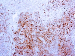
Diagnostic Biosystems MART-1/MELAN A MART-1/Melan-A, Melanoma Marker
This antibody is specific to a protein of 20 kDa known as MART-1 (melanoma antigen recognized by T cells-1) or Melan-A. This antibody does not crossreact with MAGE-1or tyrosinase. MART-1 is a newly identified melanocyte differentiation antigen recognized by autologous cytotoxic T lymphocytes. This antibody stains melanomas and other tumors showing melanocytic differentiation.
Enterprise Technology Solutions APC SMX3000RMX93 Smart-UPS 3000VA RM 120V Shipboard
This Smart-UPS features an output of 2700W / 3000VA, an input of 100-120V at 50/60 Hz ±3 Hz, six NEMA 5-15R outlets, three NEMA 5-20R outlets, a NEMA L5-30R outlet, a NEMA L5-30P input connection, line interactive topology, and a sine waveform. This Smart-UPS has an available SmartSlot for optional Network Management Card that enables graceful shutdown of physical servers, virtual machines, and HCI Clusters via PowerChute Network Shutdown.
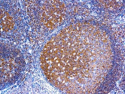
Diagnostic Biosystems BCL-10 Bcl-10
BCL10, with an N-terminal caspase recruitment domain (CARD), is found in a number of apoptotic regulatory molecules. It was identified through its direct involvement in t(1;14) of mucosa-associated lymphoid tissue (MALT) lymphoma. Expression of BCL10 was shown to induce NFĪŗB activation in a NIK-dependent pathway. This MAb labels subpopulations of normal B and T cells and is a useful tool for the sub-classification of lymphomas. In MALT lymphomas with the t(1;14) translocation, while 55% of MALT lymphomas lacking this translocation exhibited the same labeling pattern, although at a much lower level.
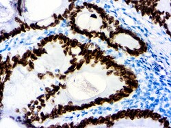
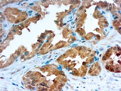
Diagnostic Biosystems SATB2 SATB2 (EP 281)
Special AT-rich sequence-binding protein 2 (SATB2) is a recently described marker that functions as a nuclear matrix-associated transcription factor. It has been reported that SATB2, in combination with CK20, could identify almost all colorectal carcinomas, including poorly differentiated colorectal carcinomas. Upper gastrointestinal (GI) carcinomas and pancreatic ductal carcinomas are usually negative for SATB2, and ovarian carcinomas, lung adenocarcinomas, and adenocarcinomas from other origin are rarely positive for SATB2. Therefore, SATB2 is a good marker for identifying a carcinoma of colorectal origin when working on a tumor of unknown primary. It can help differentiate colorectal metastasis (SATB2+) from primary pulmonary adenocarcinoma of mucinous or enteric type (SATB2- but often CDX2+)\n
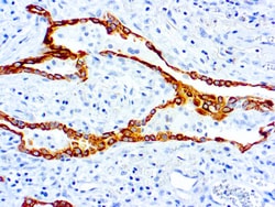
Diagnostic Biosystems CYTOKERATIN 7 Cytokeratin 7 (LP1K)
Cytokeratins comprise a diverse group of intermediate filament proteins (IFPs) that are expressed as pairs in both keratinized and non-keratinized epithelial tissue, where they constitute up to 85% of mature keratinocytes in the vertebrate epidermis. Cytokeratin 7 is expressed in epithelial cells of ovary, lung and breast. It is often used in conjunction with cytokeratin 20 and CDX-2 in distinguishing pulmonary, ovarian and breast carcinomas (CK7+) from most colon carcinomas (CK7-).
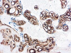
Diagnostic Biosystems COLLAGEN IV Collagen Type IV
This antibody recognizes collagen type IV epitope located on the α1 and/or α 2 chains of human collagen type IV. This antibody shows no cross-reactivity with collagen types I, II, III, V, VI and VII and does not react with human vitronectin, fibronectin or chondroitin sulfate A, B and C.
Bellco Glass, Inc. Vactrap G, 2L + 1L, Glass Bottles, Red Bin, GL45 Cap w/1/4" ID Tubingg, 1/EA
Vactrap G (Glass) is the ultimate vacuum trap system, adhering to the strictest CDC recommendations for laboratory safety. With its new continuous full vacuum rating, it is ideal for protecting vacuum lines and users. Vactrap G safely contains waste produced during supernatant removal, chemical separation, and cell or tissue culture media aspiration.Following in the footsteps of previous innovations, Vactrap G combines ease-of-use with revolutionary options and features to produce a new step forward in vacuum trap technology. From PUREGRIP glass media bottle versions to the inclusion of tangle-free VersaCap closures, to a brand new extra length tube, scientists now have access to better solutions at a lower cost.
Diagnostic Biosystems GALECTIN-3 Galectin-3
Galectins are a family of soluble β-galactoside-binding animal lectins that modulate cell-to-cell adhesion and cell-to-extracellular matrix (ECM) interactions and play a role in tumor progression, pre-mRNA splicing and apoptosis. The galectin-3 protein, also known as Mac-2, hMac-2, GALBP, CBP35 or LGALS3, contains a single carbohydrate binding domain, which binds galactose-containing glycoconjugates. Galectin-3 is expressed in colonic and intestinal epithelium, inflammatory macrophages, papillary and follicular carcinomas, neoplastic astrocytes and some B and T lymphocytes. Upregulated expression of galectin-3 is involved in cancer progression and metastasis. Galectin-3 mediates the endocytosis of β1 Integrins in a lactose-dependent manner and is associated with thyroid malignancy and Crohn’s disease. It may also be used as a marker for diagnosing casesinvolving Hurthle cell adenomas and carcinomas.
Uline Adjustable Height Stainless St
Adjustable Height Stainless Steel Worktable With Bottom Shelf - 60 X 30IN

Ace Glass, Inc. Glas-Col O Series mantle support, 1000mL, fits Ace Glass 12031-19 & 12035-19
1000ML EXTENSION SUPPORT

Chemglass Life Sciences Clamps for MAGic Platform, 1, 000mL (Max 9)

Clamps for MAGic Platform, 1,000mL (Max 9).